Overcoming Knee Osteoarthritis: The Biomechanical and Cellular Blueprint for Cartilage Regeneration
Why the 'wear-and-tear' myth is destroying joints, how cartilage actually feeds and repairs through mechanotransduction, and the clinical protocol to restore pain-free knee longevity.
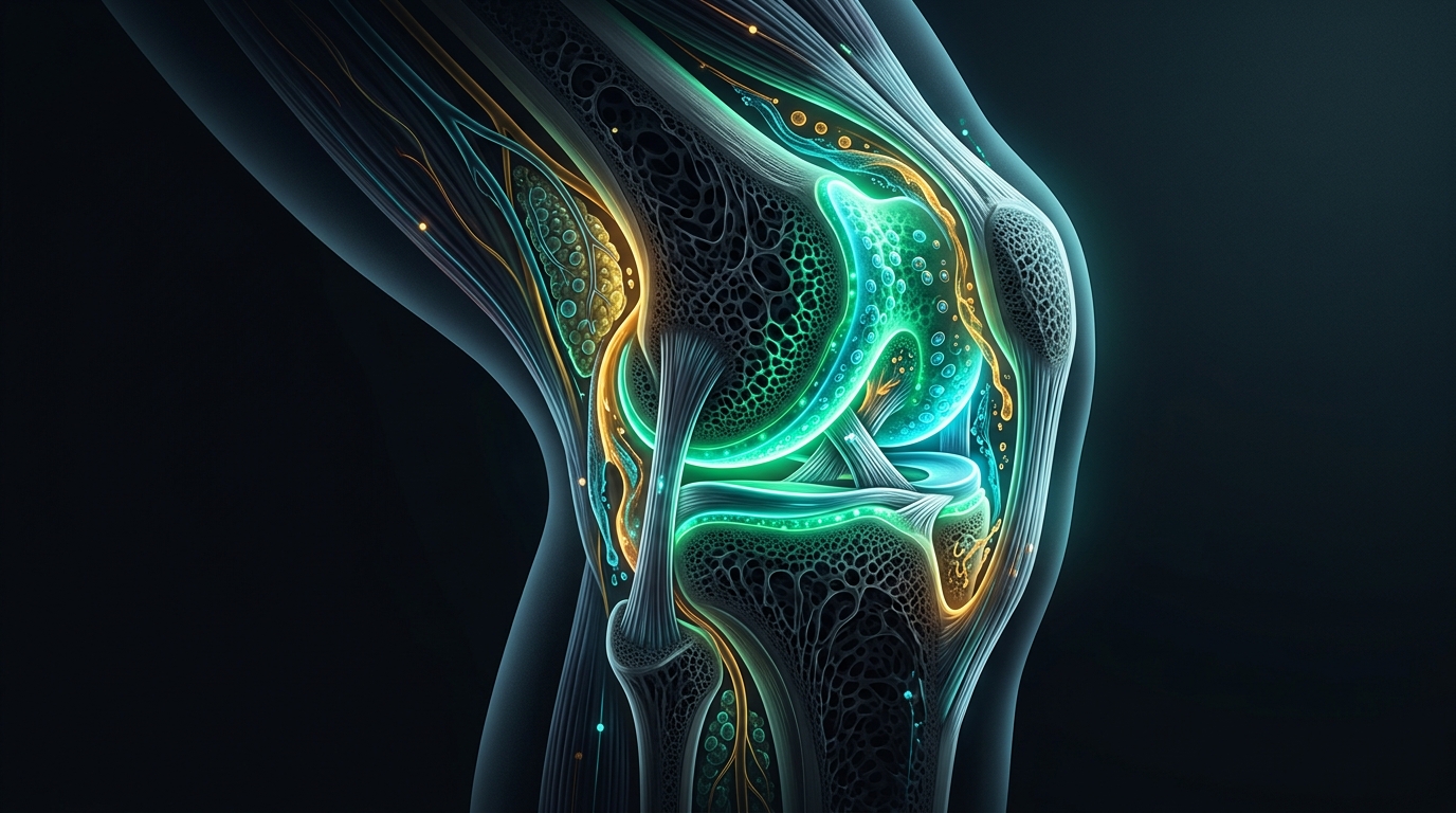
Few medical pronouncements carry the quiet finality of: "You have bone-on-bone knee osteoarthritis. Your cartilage has worn out like an old car tire. Stop running, stop squatting, avoid the stairs, and manage the pain until you are old enough for a total knee replacement."
For decades, millions of active individuals have been handed this passive death sentence for their physical autonomy. Terrified of "wearing out" what little cartilage remains, they sit down, immobilize their legs, and wait.
Yet within months of stopping movement, a tragic irony unfolds: their knees stiffen, the aching deepens, their quadriceps wither, their balance deteriorates, and walking even a single city block becomes an agonizing ordeal.
In Medicine 3.0, we recognize that the classical "wear-and-tear" model of knee osteoarthritis is not merely outdated—it is biologically backwards.
Articular cartilage is an avascular, non-innervated tissue. It possesses no blood vessels to deliver red blood cells and no direct lymphatic supply. The only way cartilage cells (chondrocytes) receive oxygen, glucose, and amino acids—and the only way they purge metabolic toxins—is through dynamic fluid imbibition driven by cyclic mechanical loading.
When you stop moving your knee, you do not protect your cartilage; you suffocate it.
Here is the cellular biochemistry, biomechanical reality, and the step-by-step clinical protocol to reclaim joint space, rebuild muscular shock absorbers, and maintain unbreakable knee longevity for life.
1. The Biological Reality: Why Cartilage is a Living Hydraulic Sponge
To heal and protect the knee joint, we must discard mechanical car analogies and look at the actual molecular architecture of articular cartilage.
The Extracellular Matrix: Type II Collagen and Aggrecan
Articular cartilage is composed of two primary functional components:
- Type II Collagen Mesh: A three-dimensional tensile grid that gives cartilage its structural shape and resists sheer forces.
- Aggrecan Proteoglycans: Giant molecular brushes studded with glycosaminoglycans (GAGs) such as chondroitin sulfate and keratan sulfate. These molecules carry dense negative electrical charges that pull water molecules inward like an ultra-powerful magnet, creating high swelling pressure.
This combination creates a hydraulic shock-absorbing cushion capable of withstanding peak loads of 3 to 5 times your body weight during walking and stair descent.
The Avascular Dilemma and Imbibition
Because cartilage has no capillaries, chondrocytes survive in an environment of physiological hypoxia.
- Compression (Loading Phase): As your foot strikes the ground, interstitial fluid is squeezed out into the joint cavity, carrying cellular waste products and inflammatory catabolites away.
- Decompression (Swing Phase): As pressure is released, the negative electrostatic charge of the aggrecan matrix rebounds, drawing fresh, nutrient-dense synovial fluid back into the tissue like water soaking into a dry sponge (fluid imbibition).
- The Sedentary Trap: If you sit all day to "save" your knees, fluid exchange halts. Chondrocytes starve, waste products accumulate, and the extracellular matrix becomes brittle, fibrillated, and prone to micro-fissuring.
2. The Paradigm Shift: Wear-and-Tear Myth vs. Modern Biological Reality
| Classical "Wear-and-Tear" Belief | Modern Medicine 3.0 Reality | Clinical Significance |
|---|---|---|
| "Cartilage wears down like a shoe sole." | Cartilage is a dynamic, living tissue that remodels under physiological cyclic load. | Total rest induces cartilage atrophy; graded loading triggers matrix regeneration. |
| "Osteoarthritis is isolated cartilage decay." | Osteoarthritis is a whole-joint organ disease involving subchondral bone, synovium, infrapatellar fat pad, and neuromuscular control. | Treating cartilage alone fails; we must address bone density, inflammation, and muscular shock absorption. |
| "Pain correlates with X-ray joint space." | Up to 40% of people with severe radiographic OA report zero pain, while many with mild X-rays suffer intense pain. | Cartilage has no nociceptive nerve fibers. Pain originates from the synovium, periosteum, bone marrow lesions, and sensitized nervous pathways. |
| "Bone spurs (osteophytes) are harmful growths." | Osteophytes are the body's compensatory attempt to widen joint surface area to stabilize an unstable joint. | Stabilizing the joint with muscular strength halts progressive osteophyte expansion. |
| "Knee pain requires stopping knee-over-toe loading." | Avoiding knee flexion creates profound weakness in the quadriceps tendon and patellar tracking. | Carefully calibrated knee-over-toe loading restores patellofemoral cartilage resilience. |
3. The Whole-Joint Disease: Why It Isn't Just Cartilage
In Medicine 3.0, the knee is viewed as an interconnected biological ecosystem. When osteoarthritis manifests, four distinct structures participate in the pathology:
🧬 The 4 Components of Knee Joint Failure
- Catabolic Molecular Cascade (MMPs & ADAMTS): Low-grade systemic inflammation (elevated IL-1β, TNF-α, and IL-6 from visceral fat) activates matrix-degrading enzymes: Matrix Metalloproteinase-13 (MMP-13) digests Type II collagen, while ADAMTS-4 and ADAMTS-5 chop aggrecan proteoglycans, destroying the cartilage's water-holding capacity.
- Subchondral Bone Remodeling & Marrow Lesions: When cartilage thins, abnormal mechanical stresses transfer directly into the subchondral bone plate. The bone reacts with microfractures, vascular congestion, bone marrow edema, and bony sclerosis. This subchondral bone is rich in nociceptive nerve endings and is a primary source of deep, aching arthritic pain.
- Synovial Inflammation (Synovitis): Cartilage breakdown fragments shed into the joint capsule, where synovial macrophages mistake them for foreign debris. The synovial membrane becomes inflamed, thickened, and produces degraded synovial fluid that loses its lubricating lubricin and hyaluronic acid concentration.
- Arthrogenic Muscle Inhibition (AMI): When the knee joint capsule experiences swelling and pain, the central nervous system reflexively shuts down neural drive to the quadriceps—specifically the Vastus Medialis Oblique (VMO). Without the quads acting as muscular decelerators, every step slams 100% of ground reaction forces directly into bone and cartilage.
4. Diagnostic Stratification: Kellgren-Lawrence Grading Scale
To design a precise rehabilitation blueprint, clinical staging using the Kellgren-Lawrence (KL) Grading System must be paired with functional assessments:
| KL Grade | Radiographic Features | Functional Symptoms | Primary Clinical Target |
|---|---|---|---|
| Grade 0 (Normal) | No joint space narrowing or osteophytes | Full painless mobility | Primary prevention, strength reserve |
| Grade 1 (Doubtful) | Minute osteophyte formation, doubtful significance | Occasional morning stiffness (< 15 min), post-exercise dull ache | Biomechanical correction, hip abductor strengthening |
| Grade 2 (Minimal) | Definite small osteophytes, possible joint space narrowing | Pain with deep squats, stairs, prolonged sitting (moviegoer's sign) | Overcoming AMI, isometrics, retro-walking, collagen peptides |
| Grade 3 (Moderate) | Moderate multiple osteophytes, definite joint space narrowing | Recurrent joint effusion, crepitus, walking tolerance < 30 minutes | Unloaded mechanotransduction, Spanish squats, hyaluronic acid support |
| Grade 4 (Severe) | Large osteophytes, severe narrowing, subchondral bone sclerosis | Persistent resting pain, severe flexion contracture, angular deformity | Pain modulation, offloading braces, surgical evaluation if conservative therapy fails |
5. The 4-Phase Joint Longevity Protocol: From Decompression to Unbreakable Strength
This evidence-based protocol breaks the vicious cycle of pain, arthrogenic muscle shutdown, and cartilage starvation:
| Phase | Core Objective | Primary Clinical Interventions | Target Dosage |
|---|---|---|---|
| Phase 1: Pain Modulation & Imbibition | De-threaten nervous system & cycle synovial fluid | Spanish Squat Isometrics, Retro-Walking (backward walk), unloaded cycling | Daily (Spanish squats: 4x45s; Retro-walk: 10–15 min) |
| Phase 2: Full Lockout & VMO Re-Education | Restore terminal extension & patellar alignment | Banded Terminal Knee Extensions (TKE), Poliquin Step-Ups, Tibialis Raises | 3 sessions/week (3x15–20 reps) |
| Phase 3: Posterior Chain & Hip Deceleration | Absorb impact before forces reach cartilage | Romanian Deadlifts, Banded Clamshells, Reverse Sled Drag | 2–3 sessions/week (3x8–12 reps) |
| Phase 4: Extracellular Matrix Nutrition | Supply rate-limiting substrates for chondrocytes | Bioactive Collagen Peptides (FORTIGEL®), UC-II®, Curcumin, Boswellia, Omega-3 | Daily nutritional protocol |
Phase 1: Pain Modulation, Joint Decompression & Imbibition
Before loading weakened cartilage, you must quiet synovial inflammation and restore neural drive to inhibited muscle fibers:
- Isometric Spanish Squat / Wall Sit Hold:
- Mechanism: Isometric contractions at 45° to 60° knee flexion trigger a profound cortical analgesic effect through local neurovascular modulation without shear friction across worn cartilage.
- Dose: 4 to 5 sets of 45-second holds, resting 2 minutes between sets, performed daily or prior to exercise.
- Retro-Walking (Backward Walking on Treadmill or Incline):
- Mechanism: Backward walking eliminates patellofemoral compressive peaks while dramatically increasing VMO firing and creating a gentle, rhythmic pumping of synovial fluid.
- Dose: 10 to 15 minutes of backward walking (0.8 – 1.5 mph, 2–6% incline or sled drag) 3–4 times per week.
- Low-Resistance Non-Impact Cycling (Joint Flushing):
- Mechanism: 10 minutes of spinning on a stationary bike with high seat height and zero resistance provides 800–900 unloaded cycles of cartilage imbibition.
Phase 2: Terminal Knee Extension (TKE) & Full Lockout Mechanics
Many arthritic knees suffer from a loss of the final 3° to 5° of terminal extension. Without full mechanical lockout, your quadriceps remain under continuous, fatiguing isometric strain during every single step:
- Banded Terminal Knee Extensions (TKE):
- Loop a heavy resistance band behind the knee, anchored forward. Step back until tension is taut. Flex the knee slightly, then contract the quad to push the knee into full lockout against band resistance.
- Dose: 3 sets of 20 controlled repetitions per leg.
- Poliquin / Peterson Step-Ups:
- Stand on a 2- to 4-inch block with the heel elevated on a wedge. Slowly lower the opposite heel to lightly tap the floor, forcing the working leg's VMO to control the eccentric knee-over-toe movement.
- Dose: 3 sets of 12–15 reps per leg.
- Tibialis Anterior Raises:
- Strengthens the front of the shin to act as the first line of ground reaction deceleration during heel strike.
- Dose: 3 sets of 20 reps leaning against a wall.
Phase 3: Posterior Chain Armor & Hip Abductor Stabilization
The knee is a "dumb joint" trapped between the foot below and the hip above. If the hip abductors (Gluteus Medius) are weak, the femur collapses inward (knee valgus), multiplying lateral and patellofemoral compressive stress tenfold:
- Side-Lying Clamshells and Standing Hip Abductions:
- Restores coronal plane stability to prevent knee collapse during walking and stair descent.
- Dose: 3 sets of 15 reps with resistance band above knees.
- Romanian Deadlifts (RDLs) & Glute Bridges:
- Strengthens the hamstrings and gluteus maximus, which serve as physical shock absorbers, pulling the tibia posterior to protect the anterior cruciate and joint space.
- Dose: 3–4 sets of 8–10 reps.
- Reverse Sled Drags:
- The single most potent restorative tool in modern joint rehabilitation. Dragging a weighted sled backwards creates continuous quad loading through full extension with zero spinal compression and minimal shear stress.
Phase 4: Targeted Cartilage Matrix Nutrition & Anti-Inflammatory Signaling
Supplements cannot fix broken biomechanics, but when paired with mechanotransductive exercise, they supply the rate-limiting molecular substrates required to rebuild the extracellular matrix:
| Nutrient / Compound | Target Daily Dose | Primary Cellular Mechanism |
|---|---|---|
| Bioactive Collagen Peptides (FORTIGEL® or Hydrolyzed Type II) | 10 – 15 g/day (taken 45–60 min before exercise with 50 mg Vitamin C) | Stimulates chondrocyte synthesis of Type II collagen and aggrecan via circulation of Pro-Hyp and Hyp-Gly dipeptides. |
| Undenatured Type II Collagen (UC-II®) | 40 mg/day (taken on empty stomach) | Induces oral tolerance in gut-associated lymphoid tissue (Peyer's patches), deactivating killer T-cell autoimmune attacks on articular cartilage. |
| Standardized Curcumin (Curcuma longa with Piperine or BCM-95) | 1,000 – 1,500 mg/day | Suppresses NF-κB transcription, directly downregulating MMP-13, ADAMTS-5, and IL-1β in inflamed synovium. |
| Boswellia Serrata (AKBA standardized to ≥ 20%) | 100 – 250 mg/day | Potent inhibitor of 5-Lipoxygenase (5-LOX), reducing leukotriene B4 and joint effusion within 7 to 14 days. |
| High-Concentration Omega-3 (EPA / DHA) | 2,000 – 3,000 mg/day (EPA > 1,500 mg) | Replaces arachidonic acid in synovial cell membranes, producing anti-inflammatory resolvins and protectins (SPMs). |
6. The Complete Weekly Joint Longevity Blueprint
| Day | Focus | Target Exercises & Dose |
|---|---|---|
| Monday | Decompression & VMO Re-Education | Spanish Squat Isometrics (4x45s) + Banded TKEs (3x20) + Retro-Walking (12 min) |
| Tuesday | Low-Impact Imbibition & Hip Stability | Stationary Cycling (15 min easy spin) + Banded Clamshells (3x15) + Glute Bridges (3x12) |
| Wednesday | Posterior Chain Strength & Deceleration | Romanian Deadlifts (4x8) + Tibialis Raises (3x20) + Farmer's Carries (3x50m) |
| Thursday | Active Joint Flushing & Core Balance | Retro-Walking (15 min on 4% incline) + Single-Leg Balance on Foam Pad (3x45s) |
| Friday | Quad Hypertrophy & Eccentric Control | Poliquin Step-Ups (3x12/leg) + Reverse Sled Drag (4x25m) + Spanish Squat (3x45s) |
| Saturday | Outdoor Zone 2 & Multi-Planar Mobility | Gentle trail hiking with trekking poles or swimming (freestyle / gentle flutter kick) |
| Sunday | Active Recovery & Cellular Renewal | Hot sauna session + light joint mobility stretching + collagen-rich nutrient feast |
Key Takeaways: Actionable Knee Longevity Blueprint
- 💡 Cartilage does not wear down like a tire: Articular cartilage is an avascular sponge that requires dynamic cyclic loading to pump synovial fluid, oxygen, and nutrients into its matrix. Total rest starves chondrocytes and accelerates joint decay.
- 💡 X-Rays do not equal pain: Up to 40% of people with severe radiographic joint space narrowing live completely pain-free. Arthritic pain arises from subchondral bone congestion, inflamed synovium, and arthrogenic muscle inhibition—all of which are highly modifiable through targeted movement.
- 💡 Master the VMO and Terminal Extension: Restoring full knee lockout with Banded TKEs and backward walking (retro-walking) offloads patellofemoral friction and re-awakens the quadriceps to absorb daily impact forces.
- 💡 Strengthen the Hips to Save the Knees: Weak gluteus medius muscles allow the femur to cave inward during gait, concentrating body weight onto a tiny point of cartilage. Strong hips create stable alignment.
- 💡 Pair Mechanotransduction with Matrix Nutrition: Take 10g of bioactive collagen peptides with Vitamin C 45–60 minutes before joint rehabilitation to spike circulating amino acids when fluid imbibition is at its peak.
Recommended Reading
To master the biomechanics, clinical science, and exercise protocols for lifelong joint resilience, read these essential books:
- Built from Broken: A Science-Based Guide to Healing Painful Joints, Preventing Injuries, and Rebuilding Your Body by Scott Hogan
The ultimate manual for connective tissue healing, explaining how to trigger tendon and cartilage remodeling through blood-flow restriction, eccentrics, and joint nutrition. - Outlive: The Science and Art of Longevity by Dr. Peter Attia, M.D.
Explores the Centenarian Decathlon and explains why preserving lower-extremity strength and pain-free mobility is the non-negotiable foundation for independence in your final decades. - Becoming a Supple Leopard: The Ultimate Guide to Resolving Pain, Preventing Injury, and Optimizing Athletic Performance by Dr. Kelly Starrett, DPT
A comprehensive biomechanical bible filled with diagnostic mobility tests, joint capsule mobilizations, and corrective strategies to fix knee tracking and movement dysfunction.
Peer-Reviewed Clinical Citations & Landmark Evidence
Mechanotransduction and Cartilage Homeostasis under Cyclic Compression:
Gao Y, Liu S, Huang J, et al. "The ECM-Cell Mechanism of Cartilage Mechanobiology in Osteoarthritis: A Review." Frontiers in Bioengineering and Biotechnology, 2022;10:914481.
🔗 PubMed PMID: 35847425 | DOI: 10.3389/fbioe.2022.914481Dissociation Between Radiographic Knee Osteoarthritis and Clinical Joint Symptoms:
Bedson J, Croft PR. "The concordance between knee impairment and knee pain in the general population: a systematic review." Annals of the Rheumatic Diseases, 2008 Jan;67(1):19-26.
🔗 PubMed PMID: 17526615 | DOI: 10.1136/ard.2007.072454Arthrogenic Muscle Inhibition in Knee Osteoarthritis and Neuromuscular Interventions:
Rice DA, McNair PJ. "Quadriceps arthrogenic muscle inhibition: the effects of experimental knee joint effusion on motor unit recruitment and firing rate." Arthritis Research & Therapy, 2010;12(4):R150.
🔗 PubMed PMID: 20687936 | DOI: 10.1186/ar3100Effects of Backward Walking on Pain, Functional Disability, and Quadriceps Strength in Knee OA:
Alghadir AH, Anwer S, Sarkar B, et al. "Effect of 6-week retro walking on pain, functional disability, and quadriceps muscle strength in individuals with knee osteoarthritis: a randomized controlled trial." BMC Musculoskeletal Disorders, 2019 Apr 8;20(1):159.
🔗 PubMed PMID: 30961582 | DOI: 10.1186/s12891-019-2537-9Bioactive Collagen Peptides and Type II Extracellular Matrix Regeneration:
McAlindon TE, Nuite M, Krishnan N, et al. "Change in knee osteoarthritis cartilage detected by delayed gadolinium enhanced magnetic resonance imaging following treatment with collagen hydrolysate: a randomized, controlled trial." Osteoarthritis and Cartilage, 2011 Apr;19(4):399-405.
🔗 PubMed PMID: 21251991 | DOI: 10.1016/j.joca.2011.01.001
Calculate Your Exact Zone 2 Cardio Training Range
Stop guessing your aerobic intensity. Determine your precise target heart rate range for mitochondrial biogenesis and lactate clearance using Karvonen & Tanaka formulas.
Recommended Telemetry & Gear for Aerobic Longevity & VO2 Max
Evidence-based clinical protocol equipment & biohacking tools
Polar H10 ECG-Grade Heart Rate Chest Strap
Wrist-based optical sensors suffer from motion artifacts and latency. An ECG chest strap ensures you stay strictly below the aerobic lactate threshold (LT1).
Garmin Forerunner 265 / Fenix 8 GPS Longevity Smartwatch
Elite cardiorespiratory fitness (VO2 Max) conveys the single largest risk reduction in all-cause mortality known to medicine. Garmin provides continuous, validated tracking of aerobic capacity and acute:chronic training load.
Apple Watch Series 10 / Ultra 2 Longevity & Health Watch
Apple Watch serves as a primary clinical gatekeeper, featuring FDA-cleared detection of occult Atrial Fibrillation and sleep apnea breathing disturbances that silently degrade microvascular integrity.
Actionable Longevity Checklist: Next Steps
Scientific insights have maximum power when translated into micro-habits. Check off your daily targets:
Log in Habitspan Daily Protocol (15s)
Lock your 4 biological pillars daily with zero friction and zero tedious data entry.
Calculate Your Biological Cell Age
Assess your functional longevity score against chronological birth age using Medicine 3.0 metrics.