Silent Organ Reserve: Defending Your Liver and Kidneys Against Metabolic Steatosis and Nephron Dropout
The liver and kidneys possess enormous physiological reserves, quietly absorbing decades of insult without pain. Learn how to safeguard functional nephrons and reverse hepatic steatosis before irreversible fibrosis sets in.
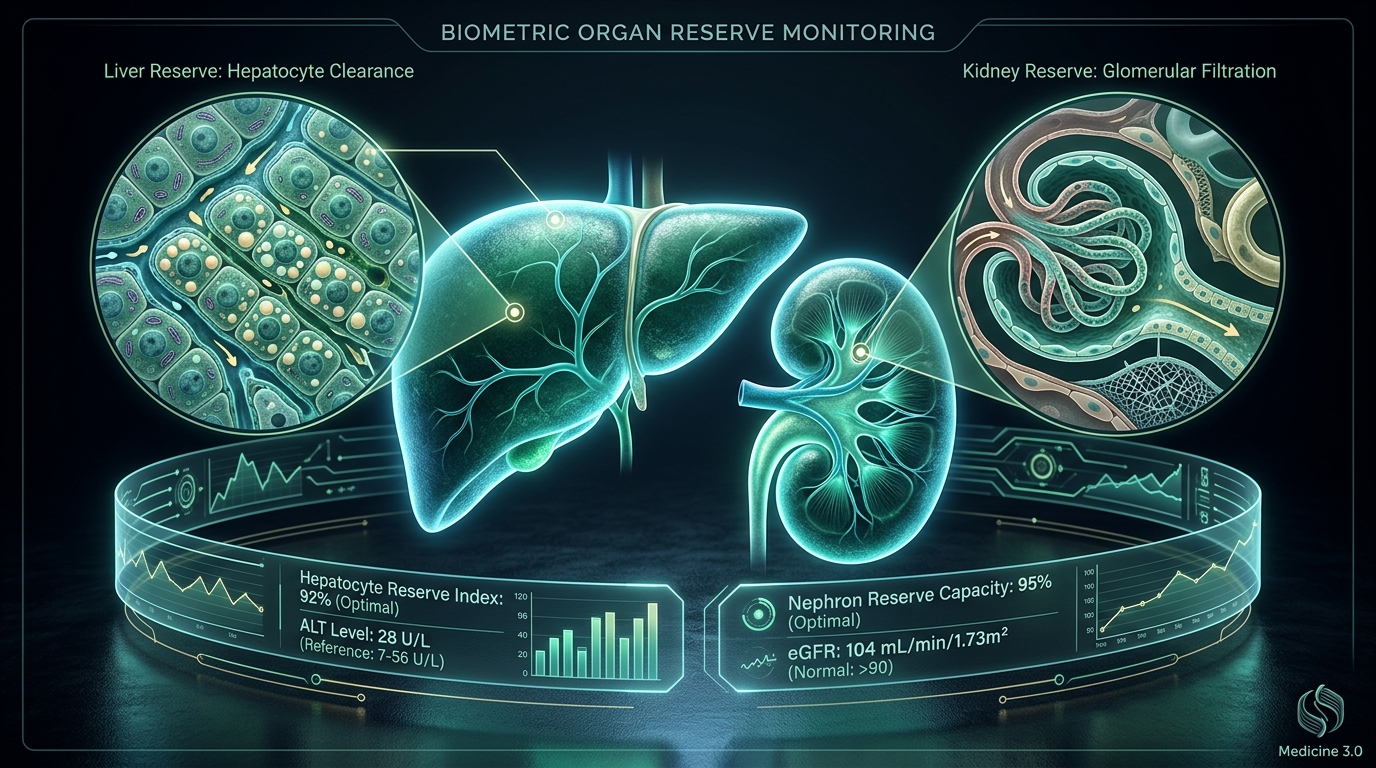
The human body is an engineering marvel designed with immense biological redundancies. Nowhere is this evolutionary buffer more profound than in our two master detoxifying and filtration engines: the liver and the kidneys.
Under ancestral conditions of famine, trauma, and infection, having surplus functional capacity meant the difference between survival and death. You can survive with only a small wedge of functioning liver tissue; you can lead a functional life with a single kidney.
However, in the modern world of ultra-processed food, ubiquitous liquid fructose, chronic psychological stress, and silent hypertension, this evolutionary blessing morphs into a deadly trap: The Illusion of Silent Organ Reserve.
Neither the liver nor the renal parenchyma possess somatic sensory pain fibers. A liver accumulating toxic triglycerides does not ache. Kidneys losing tens of thousands of microscopic filtering units (nephrons) each year do not sting or complain.
In traditional Medicine 2.0, clinical intervention is almost always delayed until overt symptoms appear—such as jaundice, ascites, peripheral edema, or severe uremic fatigue. By that stage, 70% to 80% of functional parenchymal reserve has been permanently wiped out.
In Medicine 3.0, our mission is radically proactive: we monitor cellular health markers like ALT (Alanine Aminotransferase) and eGFR (estimated Glomerular Filtration Rate) to detect subclinical organ distress decades before structural fibrosis and chronic kidney disease take hold.
1. The Liver: Deciphering ALT and the Epidemic of MASLD
The liver conducts over 500 vital metabolic biochemical reactions every single second. It governs de novo lipogenesis, glycogen storage, bile synthesis, lipoprotein assembly, and chemical clearance.
When excessive refined carbohydrates, high-fructose corn syrup, and hyperinsulinemia overwhelm hepatic mitochondrial oxidative capacity, the liver begins packaging the surplus energy into intrahepatic lipid droplets. This condition—formerly termed Nonalcoholic Fatty Liver Disease (NAFLD) and now clinically classified as Metabolic Dysfunction-Associated Steatotic Liver Disease (MASLD)—affects more than 30% of the adult global population (Younossi et al., Hepatology, PMID: 26707365).
Why "Normal" Lab ALT Ranges are Dangerous
Alanine Aminotransferase (ALT) is an intracellular enzyme primarily located within hepatocytes. When liver cells become swollen with fat droplets, undergo oxidative stress, and experience cell-membrane rupture (lipotoxicity), ALT leaks out into circulating blood.
Standard commercial laboratory reference ranges often list ALT values up to 45 to 55 U/L as "normal." This is a dangerous statistical relic. Traditional reference ranges are calculated using the 95th percentile of the population—a population that is now overwhelmingly sedentary, metabolically compromised, and steatotic.
Rigorous histological biopsy studies demonstrate that:
- True physiological "healthspan" liver targets for ALT are strictly under 25 U/L for men and under 19 U/L for women.
- An ALT of 38 U/L, while labeled "green/normal" by conventional labs, frequently conceals stage 1 or 2 steatohepatitis (MASH), actively laying down collagen and initiating hepatic fibrogenesis.
2. The Kidneys: Preserving eGFR and Halting Nephron Dropout
Each human kidney begins life with approximately 800,000 to 1.2 million filtering units called nephrons. Within each nephron, a delicate, microvascular tuft called the glomerulus filters metabolic toxins, urea, and excess electrolytes from the blood under immense mechanical pressure.
Unlike skin or liver hepatocytes, mature mammalian nephrons have virtually zero capacity to regenerate once destroyed. Once a nephron dies, its capillary tuft collapses, undergoes glomerulosclerosis, and is replaced by a non-functional fibrous scar.
The Dynamics of eGFR and Age-Related Nephron Dropout
The primary clinical proxy for functioning renal mass is the Estimated Glomerular Filtration Rate (eGFR), calculated from serum creatinine and/or cystatin C using the CKD-EPI equation (Levey et al., Ann Intern Med, PMID: 19414839, DOI: 10.7326/0003-4819-150-9-200905050-00006).
- Young, Healthy Adult Baseline: 100 – 120 mL/min/1.73m².
- The Normal Aging Curve: After age 35–40, natural physiological senescence causes an average loss of 0.8 to 1.0 mL/min/year.
- The Pathological Acceleration: In individuals with chronic subclinical hypertension (> 120/80 mmHg), insulin resistance, hyperuricemia, or chronic NSAID use, nephron destruction accelerates up to 3 to 5 mL/min/year.
By maintaining an optimal eGFR above 90 mL/min/1.73m², you preserve critical buffer capacity, ensuring your vascular endothelium, renin-angiotensin-aldosterone axis, and bone-mineral metabolism remain resilient through age 100.
3. The Modern Threats to Hepatorenal Reserve
Threat A: Liquid Fructose & De Novo Lipogenesis
Unlike glucose, which can be metabolized and stored as glycogen by every cell in the human body, fructose is almost exclusively processed by the liver. When consumed in liquid form (soda, fruit juices, sweetened teas), fructose floods the portal vein at supersonic speed. The hepatic enzyme fructokinase phosphorylates fructose without negative feedback, depleting ATP and forcing the liver into aggressive de novo lipogenesis (fat production) and uric acid synthesis.
Threat B: Subclinical Nocturnal Hypertension
The kidneys contain the most delicate microvascular capillary beds in human anatomy. When nighttime blood pressure fails to "dip" by 10–20% during deep sleep (nocturnal non-dipping), the glomeruli are hammered 24 hours a day by excessive filtration pressure (glomerular hyperfiltration), initiating microalbuminuria and progressive podocyte loss.
Threat C: Chronic Over-the-Counter NSAID Analgesics
Non-steroidal anti-inflammatory drugs (ibuprofen, naproxen) inhibit cyclooxygenase (COX) enzymes, shutting off the synthesis of vasodilatory prostaglandins (PGE2, PGI2) in the renal afferent arteriole. In periods of mild dehydration or intense athletic training, NSAID use causes acute renal medullary vasoconstriction, starving nephrons of oxygen and triggering irreversible micro-damage.
4. The 4-Pillar Clinical Protocol to Defend Organ Reserve
To safeguard your liver from steatosis and preserve functional nephron architecture, implement this tactical Medicine 3.0 protocol:
| Pillar | Protocol | |
|---|---|---|
| 1 | Nutrition | Eliminate liquid fructose, ensure 2.5–3L hydration, boost choline (egg yolks) for VLDL hepatic lipid clearance. |
| 2 | Movement | 150–180 min/week Zone 2 cardio (hepatic mitochondrial renewal). |
| 3 | Fasting | 14:10 to 16:8 circadian fasting window to trigger lipophagy. |
| 4 | Prudence | Eliminate non-essential NSAIDs; maintain blood pressure <120/80. |
1. Fuel Pillar: Eradicate Liquid Fructose & Supplement Choline
- Zero Liquid Sugars: Completely eliminate sodas, sweetened coffees, energy drinks, and concentrated fruit juices.
- Phosphatidylcholine: Consume pasture-raised eggs or sunflower lecithin. Choline is the rate-limiting nutrient required to synthesize VLDL particles to export fat out of the liver into systemic circulation, preventing intrahepatic fat trapping.
- Nephron Hydration: Maintain a daily baseline intake of 2.5 to 3.0 Liters of mineral-balanced water. Adequate volume prevents high urinary osmolarity and reduces the secretion of vasopressin (ADH), a hormone known to accelerate renal fibrosis.
2. Movement Pillar: Zone 2 Hepatic Mitochondrial Biogenesis
Engage in 150 to 180 minutes of weekly Zone 2 aerobic exercise (stationary cycling, incline walking, rowing). Aerobic muscle contractions dramatically increase whole-body insulin sensitivity, upregulate fatty acid beta-oxidation in hepatocytes, and stimulate renal capillary micro-perfusion without inducing ischemic or rhabdomyolytic kidney stress.
3. Sleep & Circadian Pillar: 14:10 Circadian Fasting
Adopt a 14:10 circadian time-restricted eating window (e.g., eat between 08:30 and 18:30). By extending the overnight fasting period, liver glycogen stores become depleted, forcing the activation of lipophagy—the selective autophagic degradation of intrahepatic lipid droplets. Furthermore, achieving 7.5 to 8.5 hours of restorative sleep restores natural nocturnal blood pressure dipping, giving the renal glomeruli vital rest.
4. Mind & Prudence Pillar: Nephrotoxic & Hepatotoxic Awareness
- Restrict NSAID use to acute emergencies only; substitute with physical therapy, heat, or targeted curcumin/omega-3 for musculoskeletal inflammation.
- Maintain strict blood pressure control (systolic < 120 mmHg, diastolic < 80 mmHg). Deep diaphragmatic breathing, sauna sessions, and stress resilience directly protect renal arteriolar tone.
Clinical Research & Evidence Citations
- Younossi, Z. M., et al. (2016). Global epidemiology of nonalcoholic fatty liver disease—Meta-analytic assessment of prevalence, incidence, and outcomes. Hepatology, 64(1), 73–84. PubMed PMID: 26707365 | DOI: 10.1002/hep.28431
- Levey, A. S., et al. (2009). A new equation to estimate glomerular filtration rate. Annals of Internal Medicine, 150(9), 604–612. PubMed PMID: 19414839 | DOI: 10.7326/0003-4819-150-9-200905050-00006
- Sanyal, A. J., et al. (2010). Pioglitazone, vitamin E, or placebo for nonalcoholic steatohepatitis (PIVENS). New England Journal of Medicine, 362(18), 1675–1685. PubMed PMID: 20427778 | DOI: 10.1056/NEJMoa0907929
- KDIGO Board. (2024). KDIGO Clinical Practice Guideline for the Evaluation and Management of Chronic Kidney Disease. Kidney International, 105(4S), S117–S314. PubMed PMID: 38490803 | DOI: 10.1016/j.kint.2023.10.018
Recommended Reading
To master metabolic optimization, establish lifelong automated health routines, and protect your internal organs from premature degeneration:
- James Clear — Atomic Habits: An Easy & Proven Way to Build Good Habits & Break Bad Ones Essential reading for eliminating liquid sugar habits, establishing consistent daily hydration, and locking in sustainable nutritional routines.
- Peter Attia, MD — Outlive: The Science and Art of Longevity A profound clinical blueprint on Medicine 3.0, metabolic steatosis, cardiovascular prevention, and proactive biomarker testing.
- Matthew Walker, PhD — Why We Sleep: Unlocking the Power of Sleep and Dreams The groundbreaking scientific guide explaining how deep sleep restores nocturnal blood pressure dipping and protects delicate organ microvasculature.
Actionable Longevity Checklist: Next Steps
Scientific insights have maximum power when translated into micro-habits. Check off your daily targets:
Log in Habitspan Daily Protocol (15s)
Lock your 4 biological pillars daily with zero friction and zero tedious data entry.
Calculate Your Biological Cell Age
Assess your functional longevity score against chronological birth age using Medicine 3.0 metrics.